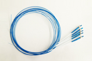
Dentine Hypersensitivity medical Laser fibers Therapy
Keywords:Dentine, Hypersensitivity, medical, fibers, Time:17-12-2015The nociceptive stimulus commonly reported in the majority of cases is that of cold, followed by the mechanical stimulus of toothbrushing and the chemical stimulus of diet with a high concentration of sugar (1). Pain of dentinal origin is sharp, located, and of short duration. The hydrodynamic theory proposed by Brännströn e Aström (2), in 1964, is still currently accepted to explain the relationship between pain of dentinal origin and the displacement of odontoblasts in the dentinal tubules. Thermal, physical and chemical stimuli would cause the displacement of the pulp-dentinal fluid, thus stimulating the pulpar nervous terminations. Under normal conditions, dentine is covered by enamel or cement and does not suffer direct stimulation. Only with the exposure of the peripheral terminations of dentinal tubules is a situation of strong dentinal sensitivity manifested, termed hypersensitivity. Occlusion of the exposed dentinal tubules can reduce the intensity of dentinal sensitivity. This can be accomplished through passive mechanisms, such as precipitation of salivary calcium phosphate inside the dentinal tubules, adsorption of plasma proteins and saliva constituents, as well by active mechanisms such as deposit of intracanalicular crystalline material and secretion of protein material from the interior of the tubules, diminishing dentinal permeability and sensitivity (3). Type A fibers are responsible for dentinal sensitivity and are probably activated by the hydrodynamic mechanism. Therefore, their activation is directly associated to the presence of opened or occluded tubules. However, hypersensitivity sometimes remains in spite of the effective blocking of the tubules, suggesting that other mechanisms contribute to nerve activation instead of or in addition to the hydrodynamic mechanism. Dentine hypersensitivity may occur as a result of sensitization induced by nerve inflammation in the dentinepulpar boundary of teeth with opened dentinal tubules. This partially explains the large sensitivity variation of exposed dentine and, furthermore, nerve activation may result in the release of neuropeptides from the activated nervous terminations and, consequently, induce neurogenic inflammation. The symptoms of dentine hypersensitivity would, up to a certain point, be self-sustainable (4). The most common factors responsible for dentine hypersensitivity are abrasion, caused by toothbrushing with inadequate intensity; abfraction, caused by tooth flexion associated with ill-directed occlusal forces, parafunctional habits or occlusal disequilibrium; erosion, as an effect of acids in the oral cavity; anatomic predisposition due to structural deficiency in the enamel-cement junction; cavity preparations in teeth with pulp vitality that expose the dentine; as well as improperly controlled dentinal acid conditioning (5,6). Any treatment, which reduces the dentinal permeability, must diminish dentinal sensitivity. The occlusion of dentinal tubules leads to reduction of dentinal permeability and, proportionally, also decreases the degree of dentinal sensitivity (7). According to the hydrodynamic theory, the effectiveness of dentine desensitization agents is directly related to their capacity to promote the sealing of the dentinal canaliculi (8). With the advent of laser fibers technology and its growing utilization in dentistry, an additional therapeutic option is available for the treatment of dentinal pain. The laser fibers, by interacting with the tissue, causes different tissue reactions, according to its active medium, wavelength and power density and to the optical properties of the target tissue (9). The laser fibers photobiomodulating action in the dental pulp was reported by Villa et al. (10), with histological studies of dental pulp of mice after irradiation with laser fibers, in teeth previously eroded with high rotation in order to expose the dentine. The profiling of the odontoblasts was observed, showing evidence of a large quantity of tertiary dentine production, causing the physiological obliteration of the dentinal tubules. The non-irradiated control teeth showed intense inflammatory process that, in some cases, evolved to necrosis. The effectiveness of dentine hypersensitivity treatment with diode laser fibers, with different wavelengths, has been reported in various clinical studies. Matsumoto et al. (11) found 85% improvement indexes in teeth treated with laser fibers; Aun et al. (12) reported successful treatment in medical fibers irradiated teeth in 98% of the cases; Yamaguchi et al. (13) reported effective improvement index of 60% in the group treated with laser fibers and only 22.2% in the control group; Kumazaki et al. (14) showed an improvement of 69.2% in the group treated with laser fibers compared to 20% in the placebo group; Gerschman et al. (15), in a double-blind study, found significant values in the treated group in relation to the placebo group: sensitivity to thermal stimuli was reduced by 67%, whereas the placebo group had a reduction of 17%, sensitivity to tactile stimuli was reduced by 65%, while the placebo group showed a reduction of 21%. The immediate analgesic effect in the treatment of dentine hypersensitivity with diode laser fibers was reported by Brugnera Júnior et al. (16) with an improvement index of 91.29% in 1102 treated teeth, operating in different bands of wavelength, 780 nm and 830 nm, and different power densities of 40 mW and 50 mW, but maintaining the same energy density deposited per dental element of 4 J/cm2. According to the consulted literature, both red and infrared wavelength laser fibers have been effective in the treatment of dentine hypersensitivity.
MATERIAL AND METHODS
A total of 40 teeth from 20 adult individuals (9 male and 11 female; aged 25 to 45 years) with a diagnosis of odontalgia of dentinal origin and cervical dentine exposure were treated. Approval by the Ethics Committee and informed written consent was obtained at the clinic of otorhinolaryngology discipline of the Escola Paulista de Medicina of UNIFESP. The teeth included in the sample were absent of bacterial infection, were at the prodromal stage of the inflammatory lesion with intense and short positive response to cold nociceptive stimulus of 0°C. The thermal test with cold stimulus was performed by the contact to the cervical dentinal surface with a flexible stick applicator, cooled with EndoFrost® (Roeko, Langenau, Germany). In order to standardize the sample, the criterion for inclusion in the study was a sensitive dentinal response of grade 10, in the 0 to 10 numeric scale for pain evaluation, characterizing cervical dentine hypersensitivity. The treated dental elements were premolars, did not have ample restorations and the individuals included in the sample showed an absence of active periodontal disease, teeth with carious lesions, chronic or debilitating disease with daily pain episodes, no use of analgesic, anticonvulsive, antihistaminic, sedative, tranquilizing or antiinflammatory medications in the 72 h preceding treatment, had not used desensitizer dentrifice in the last 3 months and had not been subjected to periodontal surgery in the last 6 months. The sample was divided into 2 groups of 20 teeth: the red laser fibers group and the infrared laser fibers group. The applied laser fibers device was the laser fibers Beam DR 500 Power, with two straight-type pens of GaAlAs diode laser fibers; one with nominal wavelength of 660 nm and verified wavelength of 660.14 nm, red, nominal power of 35 mW and verified power of 35.22 mW, focus dimension of 1 mm² with elliptical standard and the other with nominal wavelength of 830 nm and verified wavelength of 830.05 nm, infrared, nominal power of 35 mW and verified power of 35.30 mW, focus dimension of 1 mm² with elliptical standard, manufactured by laser fibers Beam Indústria e Tecnologia (Niterói, RJ, Brazil). As a study factor, the sample was also divided into 2 subgroups according age, one group consisted of 25-35-year-old individuals and the other of 35-45-yearold individuals. This study was performed by one operator and one assessor responsible for the measurement of the pain level of the patients. The treatments were carried out in 4 sessions, with intervals of 7 days between sessions, during a period of 4 consecutive weeks. The sample was evaluated through the measuring of the dentinal sensitive response to the cold nociceptive stimulus of 0ºC, with a numeric scale from zero to 10. The measurements were performed before each treatment session and at 15 and 30 min after the laser fibers application to verify the capacity, the extent, and the duration of desensitization after irradiation. This result was called immediate effect. Additional measurements were also performed at 15, 30 and 60 days after the conclusion of treatment in order to assess the extent of desensitization obtained at the different wavelengths. This result was called late effect. The dentinal cervical region of the treated teeth exposed to the buccal medium was previously cleaned.

RESULTS
Significant reduction of dentinal sensitivity occurred along all times measured during the four treatment sessions in both groups treated with red and infrared laser fibers. Comparing the means of the responses in the 4 treatment sessions of the 2 groups, the red laser fibers group showed a higher degree of desensitization in the age range of 25 to 35 years compared to the group treated with infrared Holmium laser fibers.
DISCUSSION
The literature is unanimous in demonstrating that, even with several types of treatment for dentine hypersensitivity, there is no treatment that reduces pain to satisfactory levels.
Related Articles
- Laser Fibers Application in Periodontics
- APPLICATION OF Nd–YAG LASER TREATMENT FOR ORAL LEUKOPLAKIA
- Interventional laser surgery for oral potentially malignant disorders: a longitudinal patient cohort study
- LOW LEVEL LASER THERAPY
- WHY A CO2 DENTAL LASER?
- Er:YAG laser applications in dentistry
- Study on the Influence of Semiconductor Laser Irradiated Time towards Dental Pulp and Dentin
- Laser Treatment for Failing Dental Implants
- 980 nm diode lasers in oral and facial practice: current state of the science and art
- The clinical observation of semi-conductor laser treatment of hypersensitive dentine
